Pushing the boundaries of the visible world
Washington University engineers, scientists and physicians team up to advance imaging science and improve human health
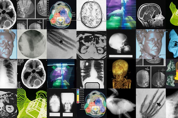
The first medical diagnostic image was taken in 1896 using an X-ray — a phenomenon discovered just a year earlier. The X-ray provided a snapshot of suspected broken bones in the wrist of a young boy. This innovation — the wild idea that doctors could see inside a body — kicked off a transformation in medicine. Since the days of those first grainy, black-and-white images, imaging scientists have created new tools and techniques to visualize the human body in ever-more-intricate detail. Today, imaging is used for everything from mapping the network of connections within the brain to diagnosing cancer to monitoring the development of a beating heart.
Washington University physicians and researchers at the School of Medicine and engineers on the Danforth Campus have been at the forefront of imaging science for more than 125 years.
The university’s first hospital was among the early leaders in adopting X-ray technology and teaching it to students. In the 1920s, Washington University researchers were the first to use X-rays with a contrast agent to view the gallbladder, making it safer and easier to diagnose gallbladder disease and paving the way for the development of contrast agents to image other organs.
In 1931, Mallinckrodt Institute of Radiology (MIR) at the School of Medicine was established to provide imaging services to support patient care and to develop the science of imaging. In the 1970s, research by Michel Ter-Pogossian, PhD, and Michael E. Phelps, PhD, at MIR led to the development of positron emission tomography (PET) and the first PET scanner. Their colleagues, Michael J. Welch, PhD, and Marcus E. Raichle, MD, developed tools and algorithms to use PET to study the brain. Welch developed an oxygen-based PET tracer to measure brain blood flow and metabolism, and Raichle captured some of the first snapshots of the brain at work. Welch also developed similar PET tracers for other parts of the body, laying the foundation for PET to emerge as a critical tool in biomedical research and clinical imaging. PET scans now are used worldwide to detect cancer, heart disease, brain disorders and other conditions. Ter-Pogossian also helped develop the first full-body computed tomography (CT) scanner, and Washington University became home to one of the first three in the world.
Read the full story here.
Click on the topics below for more stories in those areas
Back to NewsFaculty in this story