New technology offers faster, broader 3D imaging of retinas, feasibility study shows
Imaging method for retinal diseases up to 10 times faster than traditional screening, early study finds
Each year, more than 30 million people have their eyes scanned using optical coherence tomography (OCT) to detect for diseases of the retina, such as age-related macular degeneration and diabetic retinopathy. While OCT takes excellent images, it is very sensitive to any movement — even breathing — by the patient, and is limited to a specific region of the eye. A team of biomedical engineers in the McKelvey School of Engineering at Washington University in St. Louis has performed a clinical feasibility study on a new technique that is at least 10 times faster than existing OCT scanners, which creates fewer opportunities for errors from patient movement and allows for earlier detection and treatment of eye diseases.
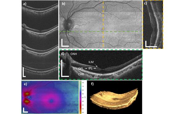
Chao Zhou, associate professor of biomedical engineering, led a team in testing a light-splitting technique known as space-division multiplexing (SDM) that takes four high-definition OCT images simultaneously with a single detector. Working with ophthalmologists at the Scheie Eye Institute at the Penn Presbyterian Medical Center at the University of Pennsylvania, Zhou and doctoral student Jason Jerwick tested the technique on 10 patients ages 18-80 with retinal disease and were able to acquire images in a wider field than existing OCT scanner in less than 1 second. The results of the feasibility study are published in Photonics Research April 1.
Because of limitations on imaging speed, the existing OCT scanners can only take an image from a limited region of the retina, which may result in missed defects or disease in the peripheral retina and an incorrect diagnosis. To address this, Zhou and Jerwick built a prototype system, which looks similar to equipment used for a regular eye exam. For the SDM-OCT scan, the patient places his or her chin on a chin rest and leans forward so that the laser beam can scan much more of the eye at a resolution of about 7 micron, or seven-millionths of a meter. The exposure has minimal risk to any damage to the eye.
The prototype used fiber-based splitters to facilitate the parallel imaging that allowed them to get four images without overlapping. Then, they stitched the images together to get a high-definition, 3D image.
"We can get similar image quality with a much higher speed and the wider field of coverage so that we can detect the peripheral disease more effectively and at an earlier stage," Zhou said.
With recent funding from Washington University's Skandalaris Center Leadership and Entrepreneurial Acceleration Program (LEAP), Zhou and Jerwick plan to rebuild the prototype SDM-OCT scanner at Washington University and will work with Rajendra Apte, MD, PhD, the Paul A. Cibis Distinguished Professor of Ophthalmology and Visual Sciences at Washington University School of Medicine, to retrofit existing OCT scanners with the SDM technology and eventually use the technology with Apte's patients to further validate its accuracy. In the new prototype, they plan to incorporate a photonic chip about the size of a quarter to split the light instead of the fiber-based splitters.
"The chip can do a lot of work for us to do the splitting and optical delay to make things happen," Zhou said.
In addition, Zhou and Jerwick are developing a high-speed handheld model that could be used with pediatric patients in collaboration with Margaret Reynolds, MD, and Andrew R. Lee, MD, both assistant professors in the Department of Ophthalmology & Visual Sciences at Washington University School of Medicine.
Jerwick J, Huang Y, Dong Z, Slaudades A, Brucker A, Zhou C. Wide-Field Ophthalmic Space-Division Multiplexing Optical Coherence Tomography. Photonics Research, 8, 537-547 (2020), published online Jan. 23, 2020, in print April 1. DOI: 10.1364/PRJ.383034.
Funding for this research was provided by National Science Foundation (DBI-1455613, 465 IIP-1623823, IIP-1640707) and the National Institutes of Health 466 (R01-EB025209).