Chan Zuckerberg Initiative names two WashU groups Frontiers of Imaging grantees
Teams were awarded $1M each to overcome limits of optical imaging
Imaging of proteins, cells and tissues is critical to understanding health and disease. Today, the Chan Zuckerberg Initiative (CZI) announced $2 million in funding for research led by faculty at Washington University in St. Louis. The support is part of nearly $32 million in funding from CZI to support biomedical imaging researchers, technology development, and the BioImaging North America international network of bioimaging facilities and communities.
Three McKelvey School of Engineering faculty members will be working with faculty from the School of Medicine and from other universities.
“We want to enable researchers everywhere to visualize, measure and analyze the biological processes underlying health and disease,” said CZI Head of Science, Cori Bargmann. “That means taking multiple approaches.”
These multiple approaches are all aimed at overcoming the same problem: imaging multiple scales of activity — including cellular and subcellular activity — in processes, in action, in situ, in real-time, without a single incision.
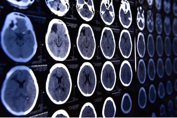
Two multidisciplinary groups headed by faculty from the McKelvey School of Engineering will be honing in on the brain, where currently, imaging techniques can penetrate about the depth of a couple of human hairs.
This is because tissue in the brain scatters and absorbs light, limiting the usefulness of microscopy. And other, non-optical imaging techniques (MRI, PET, and USI), only provide a narrow range of resolution.
“People recognized that this was a fundamental problem for classical optics,” said Jung-Tsung Shen, associate professor in the Preston M. Green Department of Electrical & Systems Engineering. He is principal investigator of a research team working with co-PIs Lihong Wang from California Institute of Technology and Junichiro Kono from Rice University.
Since classical optics had reached a barrier as far as deep-tissue imaging, Shen is focused on a different approach – exploiting the quantum entanglement between probing photons. The team is developing what might be referred to as a quantum photonic-dimer laser, a light source that produces a special class of entangled pairs of photons known as photonic dimers.
“Until now, a laser has been the only coherent, quantum light source we have; but light allows other possibilities,” he said. And so, they are looking for something different to use as the light source for imaging, in conjunction with fluorescence. Shen turned to an entirely new quantum state. “A brand-new class of coherent, quantum light,” he said.
A different approach will utilize photoacoustics. “If successfully realized, our technology will represent a quantum leap in molecular imaging, and we expect broad adoption across biomedical sciences and medicine,” said Song Hu, associate professor of biomedical engineering.
Hu will be working with co-PIs Lan Yang, the Edwin H. & Florence G. Skinner Professor in the Preston M. Green Department of Electrical & Systems Engineering and Adam Kepecs, BJC investigator and professor of neuroscience and psychiatry in the School of Medicine. Together, the team is developing a new photoacoustic technology that will enable cellular-resolution molecular imaging deep inside live tissue.
Their photoacoustic localization molecular microscopy will rely on a new, highly sensitive nanophotonic sensor, which has already achieved a thousand-fold improvement in sensitivity compared to conventional ultrasonic sensors.
In the first phase, the team expects to demonstrate an ultrasonic sensor with unparalleled sensitivity for photoacoustic imaging by exploiting the quantum physics of so-called exceptional points to record an unprecedented number of neurons across the entire mouse brain.
“Our goal is to support the advancement of imaging technologies and provide access to and training on these state-of-the-art tools so that researchers can drive towards discoveries,” said CZI Imaging Program Officer Stephani Otte. “By collaborating closely with the imaging community and providing both funding and expertise in technology development, we hope to help make the next breakthroughs in imaging possible.”
The initial funding is for 2 1/2-year pilot projects. In the second phase of the RFA, successful grantees will be eligible to apply for four-year, $10 million technology development grant awards. View the full list of grantees.
Click on the topics below for more stories in those areas
- Research
- Biomedical Engineering
- The Institute of Materials Science & Engineering
- Electrical & Systems Engineering