A disordered domain plays a key role in cell division
Research in Rohit Pappu’s lab may facilitate the design of precision drugs
Antibiotics prevent bacteria from growing and multiplying, making cell division a particularly appealing target for drug development. To illuminate the mechanisms responsible for cell division in bacteria and facilitate the design of precision drugs, biophysicists in the McKelvey School of Engineering at Washington University in St. Louis collaborated with microbiologists in the Department of Biology to discover how a small part of a key bacterial division protein functions at the molecular level.
The lead author of the study, Min Kyung Shinn, a postdoctoral fellow, and her colleagues, Megan C. Cohan, who earned a doctorate in biomedical engineering in 2020 in the laboratory of Rohit V. Pappu, the Gene K. Beare Distinguished Professor of Engineering, and Kiersten Ruff, a senior scientist in the Pappu lab, collaborated with Jessie L. Bullock, a graduate student in the laboratory of Petra A. Levin, the George William and Irene Koechig Freiberg Professor of Biology, to investigate how the intrinsically disordered region (IDR) of the essential cell division protein FtsZ actually functions. FtsZ is found in nearly all bacteria. This IDR was previously shown by Levin’s lab to be essential for cell division in the soil bacterium Bacillus subtilis.
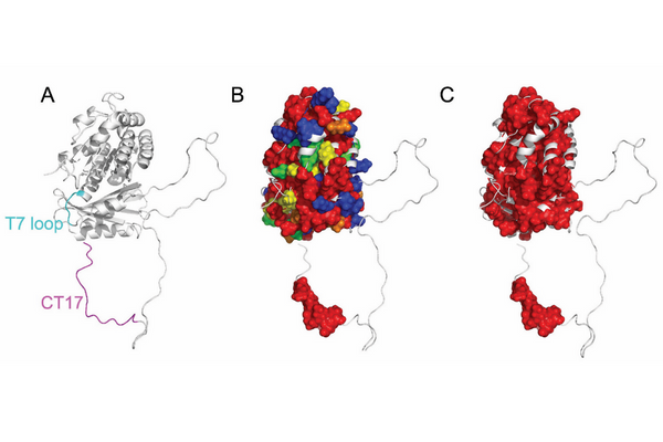
The research, which was published Oct. 10 in PNAS, builds on several key findings made over the past decade in the Pappu and Levin labs. B. subtilis FtsZ has three major parts: the protein’s folded core which is able to polymerize on its own and is nearly identical to cores from other bacterial FtsZs; the CT17 tip that acts as a molecular hub for interaction with other cell-division proteins; and the intrinsically disordered C-terminal linker that connects the core and the CT17. The Levin lab showed that the linker is essential for proper cellular localization of FtsZ and for cell division. The follow-up work of Cohan showed that the linker is an autoregulator of biochemical functions of FtsZ, but the molecular mechanisms remained unclear.
“IDRs lack distinct stable structures, and their functions are often difficult to predict,” Shinn explained. “So, we used computational simulations and discovered that the CT17 region interacts with specific regions on the core domain of FtsZ, thus revealing the molecular basis for the latchkey-like autoregulation of FtsZ’s biochemical functions.”
Shinn said that they studied more than 1,000 FtsZ protein sequences from different bacteria and found that despite the hypervariability of the linker, the interacting regions in the core and the CT17 in the molecule evolved together.
“Key sequence patterns, uncovered using novel methods that Megan Cohan and I co-developed, show that the disordered linker enables conserved functionalities in sequence-specific ways,” Shinn said.
“Every bacterial FtsZ has a distinct linker,” added Pappu, who heads the lab where the work was conducted. “This finding offers a way of thinking about how to design bespoke antibiotics to target the linker in a bacterium to stop cell division. This finding is important because it brings us closer to developing novel targets to kill disease-causing bacteria.”
“What is exciting to me is that FtsZ’s IDR, which often gets ignored because it is disordered, plays such an important role in fine tuning the function of this essential protein,” Levin said. “It is a good reminder that Mother Nature conserves things for a reason.”
The concept for the study began with Cohan’s doctoral thesis. Shinn took up the work after Cohan earned a doctorate from Washington University in 2020 and joined the Pappu lab.
“Down the line, scientists could use our findings to possibly develop an antibiotic that only targets a specific organism,” Shinn said.
Shinn MK, Cohan MC, Bullock JL, Ruff KM, Levin PA, Pappu RV. Connecting sequence features within the disordered C-terminal linker of Bacillus subtilis FtsZ to functions and bacterial cell division. PNAS, Oct. 10, 2022. https://doi.org/10.1073/pnas.2211178119
Funding for this research was provided by the Air Force Office of Scientific Research (FA9550-20-1-0241); National Science Foundation (MCB1614766) and National Institutes of Health (5R25GM127331).