Engineers develop new way to use low-count imaging for effective radiopharmaceutical therapy
Collaboration between School of Medicine, engineers in Jha’s lab opens new frontiers
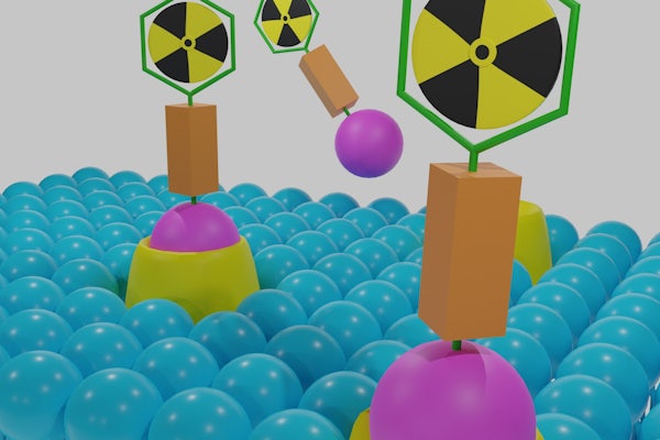
Some types of cancer and other diseases are treated with a drug containing radioactive material that targets the tumor. However, the drug can also be deposited in healthy organs, potentially damaging the kidneys, gut or bone marrow.
Abhinav Jha, a biomedical engineer in the McKelvey School of Engineering at Washington University in St. Louis, students in his lab and collaborators have developed a way to measure the distribution of alpha-particle emitting radiopharmaceutical therapy. Alpha particles are a form of radiation that have potent toxic effect on cells that is highly localized. In a variety of tests, they found that the proposed low-count quantitative single-photon emission computed tomography (LC-QSPECT) method provided reliable measurements of the radionuclide uptake.
Results of the research were published online in IEEE Transactions on Radiation and Plasma Sciences May 23, 2022.
The project began when Daniel Thorek, associate professor of radiology at the School of Medicine’s Mallinckrodt Institute of Radiology, approached Jha with a highly important need for these therapies.
“Knowing how much of the drug has gone to a certain region is needed for multiple reasons such as post-therapy management of patients who have been given this treatment,” said Jha, whose lab develops computational imaging methods to diagnose and treat diseases. “Fortunately, these drugs also emit gamma-ray photons, which can be captured by tomographic imaging systems, SPECT.”
This measured data, referred to as the projection data, can then be used to generate images of the isotope distribution inside the body, a process referred to as image reconstruction. This provides a way to quantify how much of the drug has gone where. However, the number of these photons is extremely low. This makes the reconstruction task very challenging, Jha said.
To address this issue, Jha and Zekun Li, a doctoral student in Jha’s lab, developed a method that measured uptake within different organs and tumors directly from the SPECT projection data without performing the reconstruction step. This unconventional approach to quantification made the problem much more solvable.
Jha, who also is an assistant professor of radiology at the School of Medicine’s Mallinckrodt Institute of Radiology, said that while the basic idea is not new, this work is the first to apply it to quantitative SPECT in alpha particle radiopharmaceutical therapies and is a step toward a reconstruction-less approach to quantification.
The team evaluated the efficacy of their method using multiple experiments. This included clinically realistic simulation studies, including a virtual clinical trial, in which they simulated the imaging of 50 patients with prostate cancer that had spread to the bone. The second study used a 3D-printed human-like phantom that simulated vertebrae with a lesion within the spine.
“Our goal is to measure how accurately and how precisely we have been able to measure the tracer uptake in the different regions,” Jha said. “Precision is especially important when you have such noisy data. We found that our method was not just highly accurate and outperformed conventional methods, but also highly precise,” Jha said.
This work opens new frontiers on performing calculations of the radiation dose that is obtained in these regions and can have a strong impact on this mode of treatment.
“This is an impressive technical feat that overcomes longstanding issues in low-sensitivity SPECT quantitation,” said Thorek, also a professor of biomedical engineering and radiation oncology.
Next, Jha’s lab is working with a different targeted therapy agent, thorium, a naturally occurring radioactive metal that decays to radium. Li has developed a technique to separate the two radioactive materials and measure the amount of radiation they produce in the body. In addition, they are looking to validate the measurement technique on different SPECT systems to determine if the method is reproducible across different scanners and systems.
Li Z, Benabdalla N, Abou DS, Baumann BC, Dehdashti F, Ballard DH, Liu J, Jammalamadaka U, Laforest R, Wahl RL, Thorek DLJ, Jha AK. A projection-domain low-count quantitative SPECT method for alpha-particle emitting radiopharmaceutical therapy. IEEE Transactions on Radiation and Plasma Sciences, published online May 23, 2022. DOI: 10.1109/TRPMS.2022.3175435
Funding for this research was provided by the National Institutes of Health (R21-EB024647, R01-EB031051, R56-EB028287) and the Society of Nuclear Medicine and Molecular Imaging 2020 Student Research Grant.