Brain movement measured for clues to prevent, reduce traumatic brain injury
Phil Bayly’s lab, collaborators study patterns of brain deformation in human volunteers
When the human head experiences any kind of movement — from nodding yes or no to heading a soccer ball or being jolted in a car crash — the brain moves inside the skull, leading to deformation of the tissue. Such deformations are key to understanding traumatic brain injury but are challenging to study since the brain is hidden inside the skull.
Philip V. Bayly, the Lee Hunter Distinguished Professor and chair of the Department of Mechanical Engineering & Materials Science in the McKelvey School of Engineering at Washington University in St. Louis, and Jordan D. Escarcega, a mechanical engineering doctoral student in Bayly’s lab, led a multi-institutional team to compare how the human brain deforms in response to movement using two types of magnetic resonance imaging (MRI). Their work is published in the August 2023 issue of the ASME Journal of Biomechanical Engineering.
The researchers measured the impulse response of the brain — its natural reaction to impact —and its harmonic response, or response to skull vibration at a particular frequency, similar to the experience of driving on rumble strips on the side of the road.
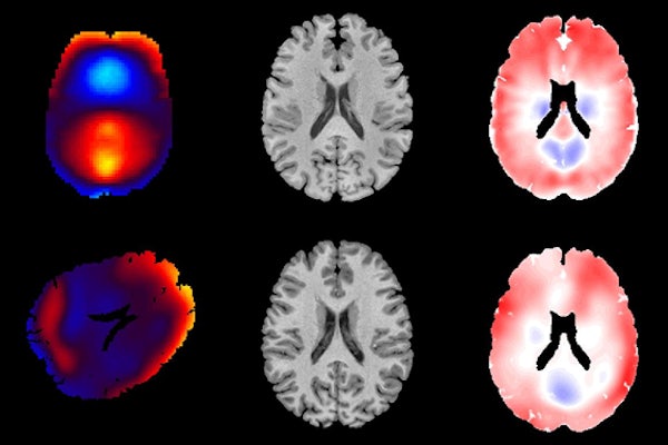
In the first case, the human volunteers’ heads were resting in a cradle while undergoing tagged MRI imaging. The volunteers were asked to gently rotate their head side to side — as if shaking their head “no” — until the cradle stopped the movement, which created the impulse response. To see the impulse response of the brain in tagged MRI, a grid of lines is superimposed over the MR image of the moving brain to see the deformation of the brain when the volunteer’s head was stopped by the cradle.
In the second experiment, light sound pressure generated by a speaker was applied to the volunteers’ heads to generate a harmonic response. Brain motion was measured using MR elastography, or MRE, a noninvasive technique that combines MRI images with low-frequency vibrations to create a visual map that shows information on body tissues.
“In the harmonic response, you’re getting a single sustained wave pattern, whereas in an impulse response, you’re getting a complex behavior that consists of the responses to multiple frequencies added together,” said Escarcega, who first came to Washington University as part of the Washington University Summer Engineering Fellowship Program (WUSEF), then returned to earn a doctorate. “Using the tagged MRI, we are able to break down the impulse response into the most dominant components and compare those to the harmonic waveforms that we excite in MRE.”
“Every human brain is different – it might as well be like your fingerprint,” Escarcega said. “But what we found was the physical patterns of deformation produced by MR elastography are extremely similar to the patterns you see from when people bump their heads.”
Tagged MRI is more difficult to perform than MRE because it requires participants to move their heads many times. Bayly and Escarcega said that MRE, which is simpler and more widely available, can be used to show the brain’s response to head impact. This result has important practical implications, they said.
“Using MRE, we can quantitatively measure patterns of brain deformation that are expected in response to head impact, but without the longer experiments and added difficulty of tagged MRI,” Bayly said. “These measurements will be useful to evaluate and improve computer models of brain biomechanics, which can then inform the design of protective equipment and reducing the societal burden of traumatic brain injury.”
###
Escarcega, JD, Knutsen AK, Alshareef AA, Johnson CL, Okamoto RL, Pham DL, Bayly PV. Comparison of Deformation Patterns Excited in the Human Brain in Vivo by Harmonic and Impulsive Skull Motion. ASME Journal of Biomechanical Engineering, August 2023, Vol. 145: 081006-1. DOI: https://doi.org/10.1115/1.4062809
Funding for this research was provided by the National Institutes of Health (U01 NS112120, R01/R56 NS05951, F31 NS122281-01A1) and the NIH Bench-to-Bedside Award in the Intramural Research Program; the Department of Defense Center for Neuroscience and Regenerative Medicine.