
Imaging tech produces real-time 3D maps of uterine contractions during labor
Noninvasive technique could shed light on preterm birth
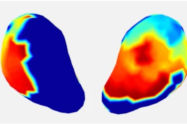
Researchers at Washington University School of Medicine in St. Louis have developed new imaging technology that can produce 3D maps showing the magnitude and distribution of uterine contractions in real time and across the entire surface of the uterus during labor. Building on imaging methods long used on the heart, this technology can image uterine contractions noninvasively and in much greater detail than currently available tools, which only indicate the presence or absence of a contraction.
The clinical study, which included 10 participants in labor through childbirth, is published March 14 in the journal Nature Communications.
“There are all kinds of obstetrics and gynecological conditions that are associated with uterine contractions, but we don’t have very accurate ways of measuring them,” said senior author Yong Wang, PhD, an associate professor of obstetrics & gynecology, of electrical & systems engineering, of radiology, and of biomedical engineering. “With this new imaging technology, we are basically upgrading the standard way of measuring labor contractions — called tocodynamometry — from one-dimensional tracing to four-dimensional mapping. This kind of information could help improve care for patients with high-risk pregnancies and identify ways to prevent preterm birth, which occurs in about 10% of pregnancies globally.”
Read the full story here.


