
Treadmill for microswimmers allows closer look at behavior
Mark Meacham leads WashU team in creating an acoustic microfluidic device to study swimming microorganisms
A team from the McKelvey School of Engineering at Washington University in St. Louis and Massachusetts Institute of Technology has created an acoustic microfluidic method that offers new opportunities to conduct experiments with swimming cells and microorganisms.
“The cells that our collaborators study are powerful swimmers for their size, and so the forces needed to trap them are substantial,” said J. Mark Meacham, associate professor of mechanical engineering & materials science in the McKelvey School of Engineering and senior author of the paper published in Proceedings of the National Academy of Sciences (PNAS) June 16, 2023. “In our devices, ultrasonic waves like those used for imaging are able to hold a cell’s body in place without affecting the way it swims.”
The cells used in the research were the single-cell alga Chlamydomonas reinhardtii, a model organism used to study the motion of cilia, tiny hairlike structures that move fluids and propel cells.
The new approach was motivated by past work in the labs of Philip Bayly, the Lee Hunter Distinguished Professor and chair of the Department of Mechanical Engineering & Materials Science who researches cilia motion, and Susan Dutcher, professor of genetics at Washington University School of Medicine, an expert in cilia structure and function, both co-authors of the paper. Meacham developed the method and devices with first author Mingyang Cui, who earned a master’s and a doctorate in mechanical engineering at McKelvey Engineering in 2017 and 2021, respectively, and is now a postdoctoral researcher at Massachusetts Institute of Technology (MIT).
C. reinhardtii cells are microscopic — and their cilia are even smaller — but they swim about 10 body lengths per second. Their cilia also beat around 60 to 70 times per second.
“The field of view at the resolution needed to see the cilia motion is such that the cells swim out and away from where you’re looking very quickly,” Meacham said. “It is difficult to study their swimming behavior without trapping the cells in some way.”
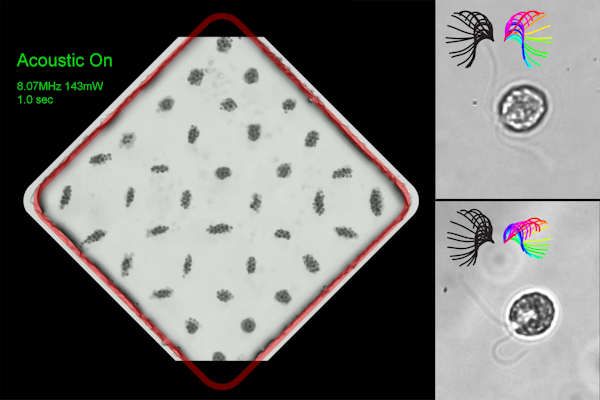
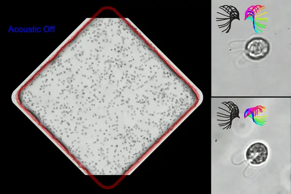
Cui overcame the trapping issue by using a combination of two types of acoustic waves. A surface acoustic wave creates vibrations that travel along the surface of a material, and a bulk acoustic wave is generated by the surface vibrations in the fluid where the cells are located.
“Cells are held by acoustic waves in the fluid at what are called nodes or low pressure regions,” Meacham said. “We wanted to use surface acoustic waves because they allow higher frequencies that give smaller traps with less distance between them, and that gives better control over the cells you’re trying to manipulate.”
Unfortunately, conventional surface acoustic wave devices are not as efficient as their bulk acoustic wave counterparts, and efficiency is needed to generate enough trapping force on these cells to hold them without the device overheating.
“Any inefficiency leads to heating, and that kills the cells,” Meacham said. “Mingyang came up with a device structure where a glass microchannel is used, which can convert surface acoustic waves into bulk acoustic waves to improve efficiency. Using glass also allows us to use high-resolution oil immersion microscopy.”
“Having solved these practical challenges, we could focus on the other advantages of acoustofluidic trapping,” Meacham said. “The key need for our collaborators was to trap these cells without constraining their rotation. Acoustic trapping enables that because it doesn’t contact the cells directly.”
Previously, to study these swimming C. reinhardtii cells, researchers used a suction pipette to hold the cell in place while the cilia were imaged. However, this does not allow the cell body to move even a little in response to cilia beating, particularly limiting rotation of the cell, which is the natural motion when it swims.
“Think of it as a treadmill for these microswimmers, and the acoustic field provides a way to hold the cell in place without affecting the cilia motion or swimming in three-dimensional space,” Meacham said.
The device also has additional benefits for experimental work with microswimmers.
“We can create 25 to 30 traps at one time and do all of the analysis of the trapped cells in parallel,” Meacham said. “You can’t do that with a micropipette—it’s not physically possible. This way you can perform measurements rapidly on a larger number of cells.”
Bayly said he is excited about the implications of this work for understanding cell motility.
“Mingyang’s results suggest that the method doesn’t affect swimming in any way, but the big impact may be in the flexibility of the approach to trap any swimming cells or microorganisms of this size range,” Bayly said. “You can now perform a number of new experiments to address unanswered biological questions by using acoustic trapping to provide a controlled environment for that experiment to take place.”
Cui M, Dutcher SK, Bayly PV, Meacham JM. Robust acoustic trapping and perturbation of single-cell microswimmers illuminate three-dimensional swimming and ciliary coordination. Proceedings of the National Academy of Sciences (PNAS) June 16, 2023. DOI: https://doi.org/10.1073/pnas.2218951120.
This research was supported by the National Science Foundation (CMMI-1633971 and CBET-1944063).
Click on the topics below for more stories in those areas
Back to NewsFaculty in this story

Philip Bayly

Mark Meacham

Susan Dutcher
Professor of genetics
Mingyang Cui
Postdoctoral researcher, MIT


