
Wearable, light-based brain-imaging tech to be commercialized with aid of NIH grant
Scientists, engineers receive small-business development grant
Figuring out what’s going on inside people’s heads typically requires huge, expensive equipment and volunteers willing to spend hours performing repetitive tasks while lying inside a narrow metal tube. Researchers at Washington University in St. Louis are working on an alternative. They are developing a cap that can be worn while moving around normally that will generate, using the power of light, high-resolution images of the brain in action. The project is supported by a Small Business Technology Transfer grant from the National Institutes of Health (NIH).
“Functional magnetic resonance imaging (fMRI) is the gold standard for imaging brain function, but fMRI is very loud and very constraining, and that limits what you can do,” said Joseph P. Culver, the Sherwood Moore Professor of Radiology at the School of Medicine’s Mallinckrodt Institute of Radiology (MIR) and the primary inventor of the technology. “Wearable brain-imaging tech would allow us to study how brain areas work together to solve specific tasks and govern behavior under naturalistic conditions.”
Culver started designing the first diffuse optical tomography (HD-DOT) instrument for imaging the brain in 2005. The technique uses LED sources that beam in infrared light from outside the head, paired with detectors that measure the light coming back out. The signals collected by each source-detector pair contain information about local brain blood flow. By placing many sources and detectors in an interlaced high-density array all around the head, the researchers can map blood dynamics — a proxy for brain activity — all over the brain. Recently, Culver and colleagues demonstrated that they could use an HD-DOT cap to detect brain signals and then decode them to figure out what a person sees.
Small Business Technology Transfer grants are designed to help small businesses bring academic innovations to market in collaboration with research institutions. This grant was awarded to EsperImage, a Washington University startup founded by Culver along with Adam Eggebrecht an associate professor of radiology at MIR, and Jason Trobaugh, and Ed Richter, both professors of practice in electrical & systems engineering at McKelvey School of Engineering. The four have worked together on HD-DOT technology for more than a decade.
The researchers envision the cap as a research tool for cognitive neuroscientists. Such scientists study how brain activity, as measured by neuroimaging systems, relates to the complex cognitive functioning of the mind. For example, scientists could use such a cap to image the brains of children as they freely talk and interact with their caretakers. This would help us learn more about how language networks in the brain develop and contribute to normal or abnormal language acquisition.
The first-generation HD-DOT devices weigh hundreds of pounds. A participant sat in a fixed chair and wore a headset tethered to a bank of electronics the size of a chest of drawers. The current prototype is 8 pounds, plus a power source that fits inside a backpack. The goal is to get the cap down to 4 pounds, which is about the weight of a football helmet.
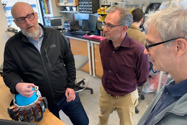
The working design calls for a cap studded with 288 optical sensors known as optodes, in the form of small copper-colored boxes about the size of an adult’s thumb. Each optode contains a light source, a detector and eight tiny circuit boards that function together as a minuscule computer. In total, 72 minicomputers are connected to a digital network through the cap, and their collective data are beamed by Wi-Fi to a central computer that acquires, analyzes and displays the data.
“The cap we have now probably took five months to build,” said Trobaugh, the co-founder and CEO of EsperImage. “The challenge now is to take the lab prototype and turn it into a commercial system that can be mass-produced and disseminated.
“This is very much a team effort that would not be possible without our four connected but different backgrounds,” he continued. “HD-DOT brain imaging was Joe’s idea originally, and Adam has helped develop and extend the technology’s capabilities, particularly toward pediatric imaging. Ed brings expertise in electrical and computer engineering, mixed analog/digital hardware and software design that has enabled him to redesign this system and build it in new ways. My background is in computational imaging science and signal processing. It takes all of our various skills and expertise to make this cap a reality.”
About Washington University School of Medicine
WashU Medicine is a global leader in academic medicine, including biomedical research, patient care and educational programs with 2,800 faculty. Its National Institutes of Health (NIH) research funding portfolio is the third largest among U.S. medical schools, has grown 52% in the last six years, and, together with institutional investment, WashU Medicine commits well over $1 billion annually to basic and clinical research innovation and training. Its faculty practice is consistently within the top five in the country, with more than 1,800 faculty physicians practicing at 65 locations and who are also the medical staffs of Barnes-Jewish and St. Louis Children’s hospitals of BJC HealthCare. WashU Medicine has a storied history in MD/PhD training, recently dedicated $100 million to scholarships and curriculum renewal for its medical students, and is home to top-notch training programs in every medical subspecialty as well as physical therapy, occupational therapy, and audiology and communications sciences.


